
文件:细胞17-29.png

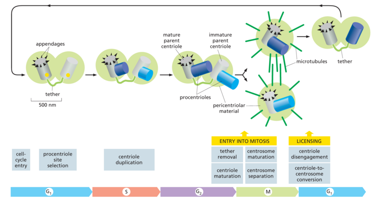
Figure 17–29 The centrosome cycle. In
early G1, the centrosome consists of two centrioles linked by a protein tether, as well as associated pericentriolar material for nucleation of microtubules (light green). One centriole, the mature parent, carries protein appendages required for certain centrosome functions. Upon entry into the cell cycle, G1/S-Cdk and Plk4 initiate centriole duplication, whereby a new centriole (procentriole) is assembled at a single site on the side of each parent centriole (yellow dots). The elongation of the procentrioles is usually completed in G2. The two centriole pairs remain close together in a single centrosomal complex until entry into mitosis, when the protein kinases M-Cdk, Plk1, and other regulators trigger numerous changes: the tether between centrosomes is removed, the immature parent centriole acquires appendages (centriole maturation), the pericentriolar material expands to enable more microtubule nucleation (centrosome maturation), and the centrosomes separate, forming a new spindle between them. After the completion of mitosis, the two centrioles in each centrosome detach (centriole disengagement), and the new centriole acquires pericentriolar material (centriole-to-centrosome conversion). These two processes are required for centriole duplication in the subsequent cell cycle; thus, progression through mitosis is required for duplication, helping to ensure that centrioles (and centrosomes) duplicate only once per cell cycle
文件历史
点击某个日期/时间查看对应时刻的文件。
| 日期/时间 | 缩略图 | 大小 | 用户 | 备注 | |
|---|---|---|---|---|---|
| 当前 | 2024年12月25日 (三) 11:06 | | 1,897 × 994(275 KB) | 长河(留言 | 贡献) |
您不可以覆盖此文件。
文件用途
以下页面使用本文件: